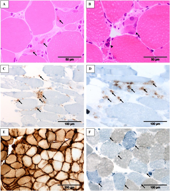
Fig 1. The myopathological features of our telbivudine-associated myopathy series.
Cytoplasmic bodies (A, arrows) within degenerate/necrotic fibers, fiber regeneration (B, arrow) and clumped nuclei (B, arrowhead), and inflammatory infiltrates consisting mainly of CD4+ T-cells (C, arrows) and CD8+ T-cells (D, arrows) as observed in Patient 4. Strong sarcolemmal overexpression of MHC class I in Patient 1 (E) and COX-negative fibers (F, arrows) in Patient 4. Stains: Hematoxylin and Eosin (A & C), immunohistochemistry with 3, 3’ diaminobenzidinetetrahydrochloride chromogen/hematoxylin (C–E) and combined COX/SDH histochemistry (F). Original magnification: x 40 objective (A & B); x 20 objective (C, D & F); x 10 objective (E).

