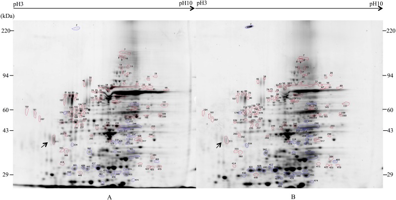
Fig 5. Two-dimensional electrophoretic analysis of hemolymph proteins from naïve and MRSA-challenged R. flavipes.
Approximately 200 μg of hemolymph proteins were separated by two-dimensional gel electrophoresis and visualized with Sypro®Ruby. Blue circles indicate protein spots that are upregulated in MRSA challenged termite and red circles indicate protein spots that are downregulated. (A) Naïve termites. (B) MRSA-challenged termites.

