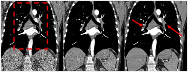
Fig 3. Comparison of FBP, iDose and IMR—coronal view at full dose.
Coronal tomographic slices of a 72-year-old male patient. The images were reconstructed with FBP, iDose and IMR (from left to right) at full dose (100% dose-level, meaning 85 mA, 100 kV and 2.25 mSv for this patient). Central and segmental pulmonary emboli can be clearly identified (arrows). The red dashed rectangle indicates the enlarged view in Fig 4. FBP = filtered back projection, iDose = iterative dose reduction, IMR = iterative model reconstruction

