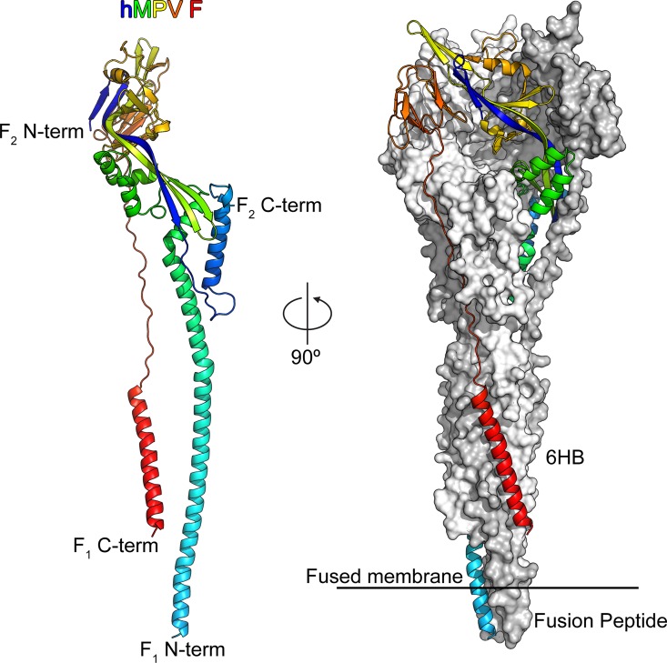
Fig 2. Structure of hMPV F in the postfusion conformation.
Left: One protomer of the postfusion hMPV F trimer is shown as a ribbon colored as a rainbow from the N-terminus of F2 (blue) to the C-terminus of F1 (red). Right: The postfusion hMPV F trimer with one protomer shown as a ribbon and two protomers shown as molecular surfaces colored white and grey. The six-helix bundle (6HB) and fusion peptides are labeled.

