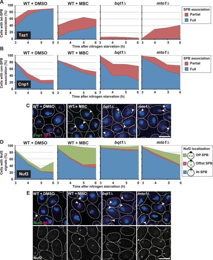
Fig 4. Effects of MBC on centromere detachment and kinetochore disassembly.
(A and B) Changes in the population of haploid cells with SPB-associated telomeres (A) or centromeres (B) during meiosis progression. SPB association of a portion of (Partial) or all (Full) Taz1/Cnp1 signals is shown by red or blue, respectively. (C) Centromere positioning in haploid cells. (D) Changes in the population of haploid cells containing Nuf2 signals during meiosis progression. Nuf2 localization is shown as in Fig 3D. (E) Nuf2 localization in haploid cells. In (C) and (E), images show cells incubated in nitrogen-free medium for 5 h. White lines show cell outlines, and arrowheads show Cnp1 or Nuf2 signals co-localized with SPB signals. Bars: 5 μm. WT+DMSO: wild-type haploid cells treated with DMSO; WT+MBC: wild-type haploid cells treated with MBC.

