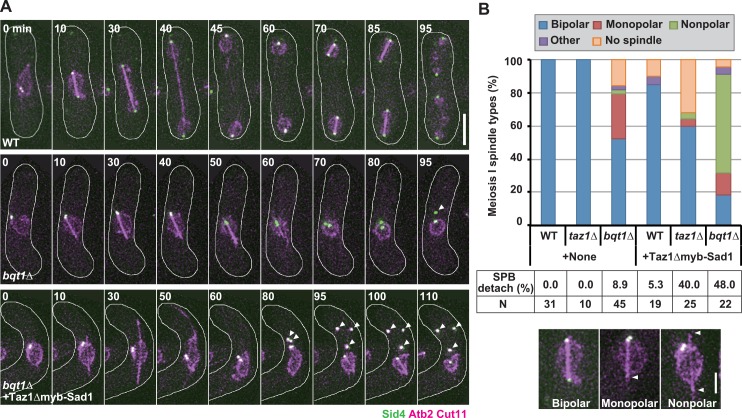
Fig 9. Effects of Taz1Δmyb-Sad1 on spindle formation in telomere clustering-defective cells.
(A) Dynamics of meiotic spindles and the SPB. Microtubules, the SPB, and the nuclear periphery were visualized by mCherry-tagged α-tubulin (Atb2), a GFP-tagged SPB component Sid4, and an mCherry-tagged nuclear pore component Cut11 [59], respectively. Images were taken every 5 min. Arrowheads indicate SPBs that are dissociated from the nuclear periphery. White lines indicate cell outlines, and numbers indicate time in minutes. +Taz1Δmyb-Sad1: with Taz1Δmyb-Sad1 expression. Bar: 5 μm. (B) Observation frequencies of various meiosis-I spindle types and SPB detachment from the nuclear periphery. +None: without Taz1Δmyb-Sad1 expression; +Taz1Δmyb-Sad1: with Taz1Δmyb-Sad1 expression. Bipolar: a normal spindle with SPBs at both poles; Monopolar: a spindle with the SPB at one of the poles; Nonpolar: an SPB-lacking spindle; Other: other types of spindles; No spindle: no spindle formation; SPB detach: percentages of cells with the SPB detached from the nuclear periphery; N: number of examined cells. Typical images of the observed spindles are shown at the bottom, and arrowheads indicate the spindle pole lacking a SPB. Bar: 2 μm.

