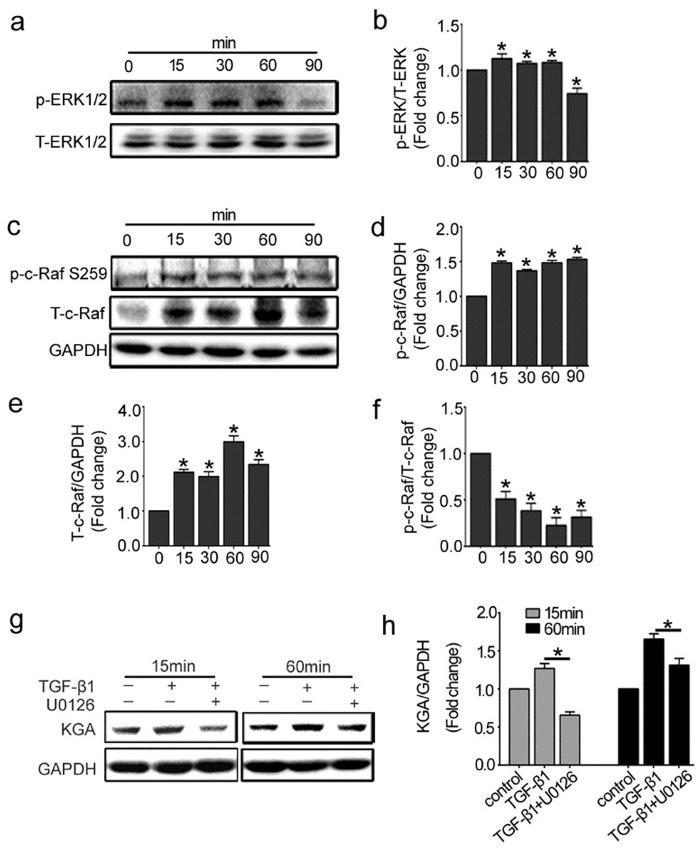
Fig 2. Raf-MEK-ERK signaling pathway is involved in KGA activation in HUVECs.
The cells were treated with 10ng/ml of TGF-β1 for different indicated periods, from 15 minutes to 90 minutes. (a, c) Representative western blot of p-ERK1/2, T-ERK1/2, p-c-Raf (Ser 259) and T-c-Raf in each group was shown. (b, d, e, f) The histogram are normalized to a GAPDH control and showed the ratio of p-ERK1/2 to T-ERK1/2 and p-c-Raf to T-c-Raf (n≥3). (g, h) HUVECs were pretreated with MEK 1/2 inhibitor (U0126, 10μM) for 30 min. KGA protein expressions were detected by western blot with densitometry analysis (n≥3). *P<0.05 compared to control group. Bars represented means ±SEM.

