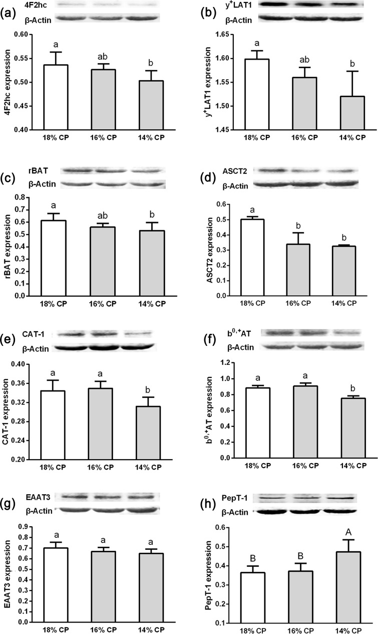
Fig 2.
Western blot analysis of the proteins 4F2hc (a), y+LAT1(b), rBAT (c), ASCT2(d), CAT-1 (e), b0,+AT (f), EAAT3 (g), and PepT-1 (h) obtained from the mid-jejunum of growing gilts fed 18%, 16% or 14% crude protein diets for 4-weeks (n = 6). β-actin was used as an internal standard to normalize the signal. One treatment included six replications, and one replication had three repeated measures. Means without a common letters differ significantly (P < 0.05).

