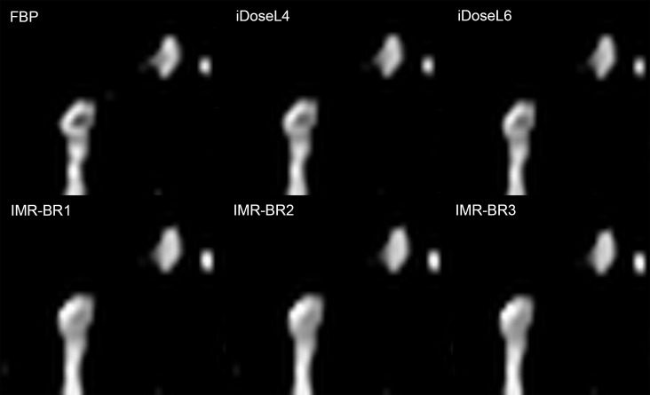
Fig 8. Conspicuity of subsegmental pulmonary embolism in RD-CTPA being reconstructed FBP, iDose4 and IMR.
Detailed enlargement of a subsegmental pulmonary artery with embolus. RD-CTPA image reconstructed with FBP, iDoseL4, iDoseL6, IMR-BR1, IMR-BR2 and IMR -BR3. Whereas the filling defect is well definable in FBP images, it’s conspicuity slightly decreases with iDose4 and markedly decreases with IMR.

