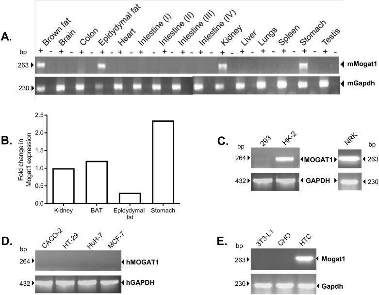
Fig 1. Expression of Mogat1 in various mouse tissues and cell lines.
A. Mogat1 expression in several mouse tissues. Mogat1 is expressed in brown adipose tissue, epididymal fat, kidney and stomach. B. Mogat1 expression was measured by RT-qPCR in the brown adipose tissue (BAT), epidydymal fat, kidney and stomach obtained from WT mice. The data were normalized to cyclophilin and then expressed as a fold change increase in Mogat1 compared to kidney. Stomach was found to be the highest expressing tissue. C. MOGAT1 is not expressed in human embroyonic kidney (HEK-293) cells but is expressed in human proximal kidney tubule (HK-2) cells and normal rat kidney cells (NRK). D. Other human cell lines screened for MOGAT1 expression: Caco-2, HT-29, Huh-7 and MCF-7. E. Expression of Mogat1 in rodent cell lines: mouse fibroblasts, 3T3-L1; Chinese hamster ovary (CHO) cells; Rat hepatic tumor cells, HTC. Mogat1 was expressed only in HTC cells. Shown is the Mogat1 product as analyzed on 1.5% agarose gels and expression of the housekeeping gene glyceradehyde phosphate dehydrogenase (Gapdh). (+) = cDNA amplification in the presences of reverse transcriptase; (-) = cDNA amplification in the absence of reverse transcriptase.

