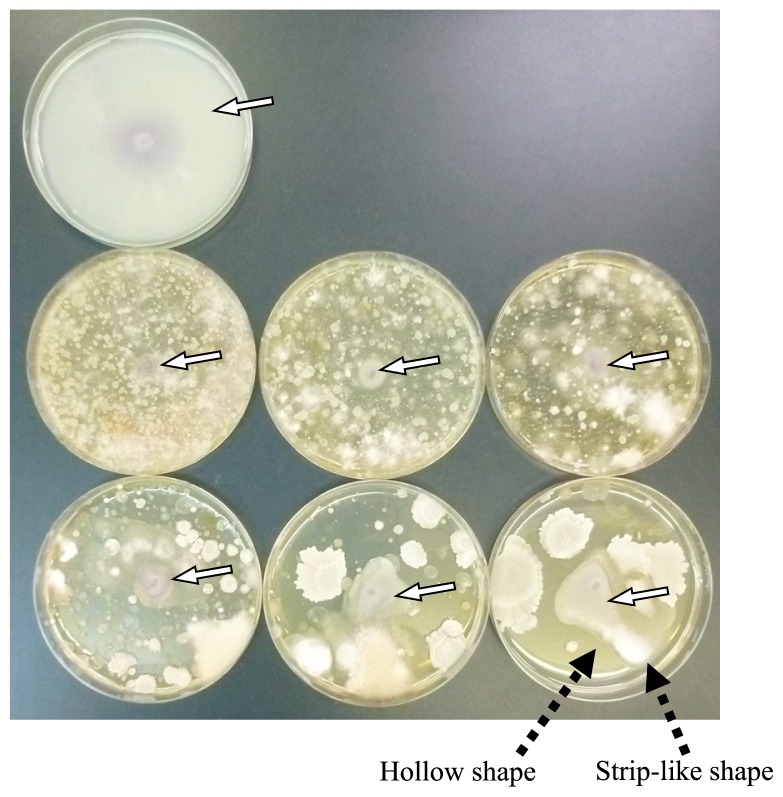
Fig. 2.
Colonies of Fusarium oxysporum f. sp. spinaciae on agar plates. A plate in the upper line, uninoculated; plates in the middle line, dilutions of 10−1, 10−2, and 10−3 from left to right; plates in the lower line, dilutions of 10−4, 10−5, and 10−6 from left to right. White arrows show colonies of F. oxysporum f. sp. spinaciae and black arrows indicate the strip-like shape and hollow shape of the colony.

