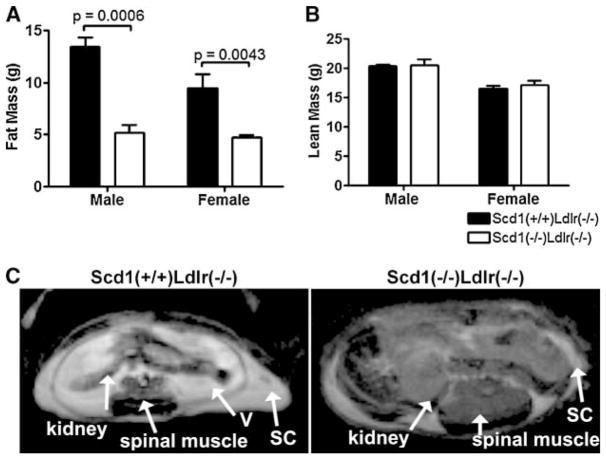
Fig. 2.
Adiposity in Ldlr−/− mice lacking SCD1. A, B: Total body fat mass (A) and total body lean mass (B) from mice fed a Western diet. n = 3–10 mice per group. Data shown are means ± SEM. C: Transverse abdominal cross-sections of an SCD1-deficient mouse and a control mouse obtained by magnetic resonance imaging. Slices (1.5 mm thick) at the kidneys were identified in sagittal images from each mouse. Fatty tissues are show as bright areas. Spinal muscle, kidneys, and subcutaneous (SC) and visceral (V) fat are indicated.

