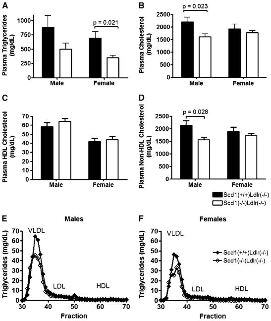
Fig. 4.
Plasma lipids and lipoprotein profiles in Ldlr−/− mice lacking SCD1. A–D: Plasma TG (A), TC (B), HDL-cholesterol (C), and non-HDL-cholesterol (D) contents of Scd1+/+Ldlr−/− and Scd1−/−Ldlr−/− mice fed a Western diet. n = 8–12 mice per group. Data shown are means ± SEM. E, F: Fast-protein liquid chromatography lipoprotein profiles of pooled plasma samples from Scd1+/+Ldlr−/− and Scd1−/−Ldlr−/− mice fed a Western diet. TG levels were determined for each fraction from male (E) and female (F) mice. The lipoprotein peaks for VLDL, LDL, and HDL are indicated.

