Figure 5.

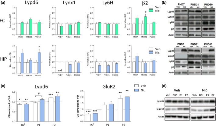
Perinatal nicotine exposure increases hippocampal Lypd6 levels. (a) Quantification of Lypd6, Lynx1, and Ly6H protein levels as well as the β2 nAChR subunit from frontal cortex (FC) and hippocampus (HIP) of male rats exposed to nicotine from embryonic day 7 to post‐natal day (PND) 21 through osmotic minipumps implanted into pregnant dams. Animals were killed at PND7 (n = 5), 21 (n = 10), or 60 (n = 13). Main effect of nicotine treatment in a two‐way anova with age and treatment as the fixed factors is indicated in the graphs. *p < 0.05 indicates statistical difference in a Sidak‐corrected multiple comparisons test. (b) Representative western blot images of protein levels from FC and HIP at the different ages. (c) Quantification of Lypd6 and GluR2 protein levels in hippocampal tissue from the P60 groups described (a), showing that perinatal nicotine exposure increases hippocampal Lypd6 levels in nuclear/endosomal fractions. Tissue was fractionated using differential centrifugation into a pellet 1 (P1, nuclear) and pellet 2 (P2, crude synaptosome) fraction or cross‐linked with bis(sulfosuccinimidyl) suberate (BS 3). Data are normalized to actin levels and the level of the untreated, non‐fractionated homogenate was set to 1 (dashed line). *p < 0.05, **p < 0.01, ***p < 0.001 indicates statistical difference from homogenate in a ratio paired t‐test, # p < 0.05 indicates statistical difference between vehicle‐ and nicotine‐treated groups in an unpaired t‐test. (d) Representative western blot images of protein levels shown in (c).
