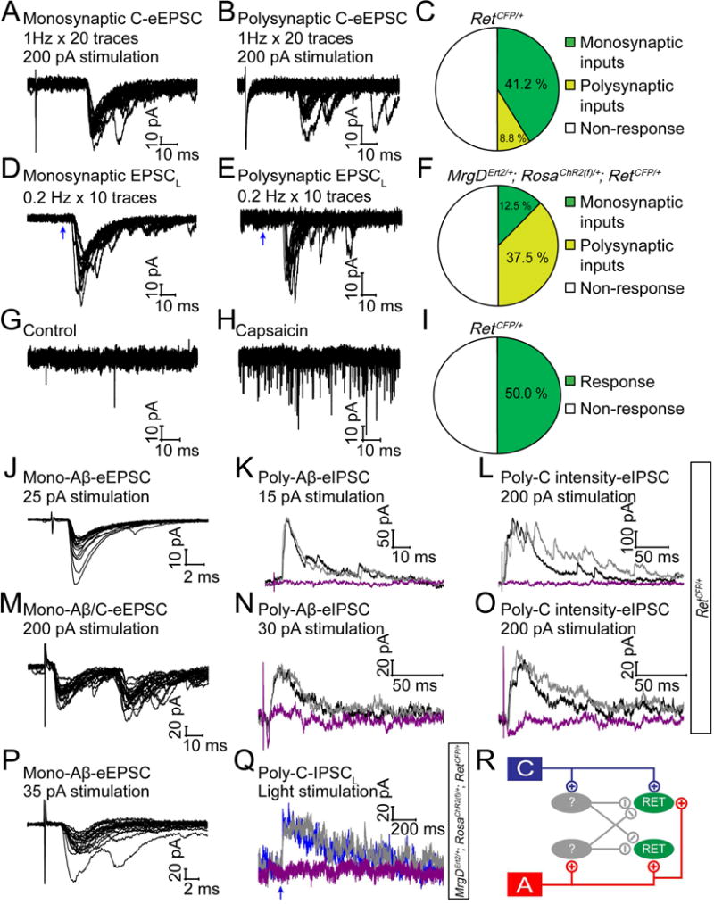
Figure 4. RET+ dDH neurons receive C fiber nociceptive inputs.

(A and B) Representative traces of mono- and polysynaptic EPSCs of RET+ dDH neurons, evoked by 1 Hz C fiber stimulation of the dorsal root. A lack of synaptic failures during 1 Hz stimulation indicated a monosynaptic response, otherwise it was considered as a polysynaptic response. (C) Quantification of C fiber induced responses. (D and E) Light-induced EPSC recording from RET+ dDH neurons in 3–5pw MrgDErt2/+; RosaChR2(f)/+; RetCFP/+mice. Mono or polysynaptic EPSCLS were differentiated by 0.2 Hz, 20 times light stimulation. (F) Quantification of light-induced responses. (G and H) 1 μM capsaicin induced responses of RET+ dDH neurons in 4–5pw RetCFP/+ transverse/sagittal spinal cord slices. (I) Quantification of capsaicin induced responses. (J–O) Two examples of RET+ dDH neurons from 3–5pw RetCFP/+mice, which received monosynaptic excitatory Aβ inputs (J, holding potential −70 mV), or monosynaptic excitatory Aβ and C inputs (M), as well as polysynaptic inhibitory inputs by dorsal root stimulation of Aβ intensity (K and N, holding potential 0 mV) or C intensity (L and O). These inhibitory inputs (black traces) were blocked by strychnine (purple trace), but not by bicuculline (gray trace). (P–Q) An example of a RET+ dDH neuron in a 3–5pw MrgDErt2/+; RosaChR2(f)/+; RetCFP/+ mouse, which received monosynaptic Aβ excitatory inputs upon dorsal root stimulation (P) as well as polysynaptic C inhibitory inputs by light stimulation (Q). The inhibitory inputs were blocked by strychnine (purple trace) but not bicuculline (gray trace) (Q). (R) Schematic showing inputs to RET+ dDH neurons.
