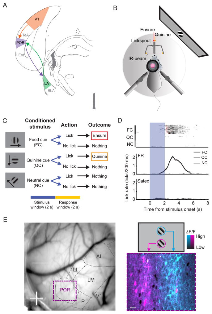
Figure 1. In vivo two-photon imaging of head-fixed mice during a Go/NoGo visual discrimination task.
A. Schematic of a V1 - POR - LA circuit.
B. Schematic of setup for in vivo two-photon imaging in a mouse performing a Go/NoGo visual discrimination task. Licking is tracked via an IR beam positioned in front of the mouse. Ensure and quinine are delivered via adjacent lickspouts.
C. The task consists of three square-wave gratings drifting in different directions. The cues are presented for 2 s, followed by a 2 s response window. If mice respond with a lick following offset of (i) the food cue (FC), they receive Ensure, (ii) the quinine cue (QC), they receive quinine, and (iii) the neutral cue (NC), they receive nothing.
D. Mice learned to lick following the FC but not following the NC or QC (top: individual licks denoted in black, Ensure delivery in red, quinine delivery in orange). Food-restricted mice performed at a high level. Following satiation, mice refrained from licking and received far fewer food rewards.
E. Left: Image of the mouse brain through a cranial window with visual areas demarcated based on intrinsic autofluorescence signal retinotopic mapping, which guided the injection of AAV-GCaMP6f into either V1 or POR. Further retinotopic mapping was done using two-photon calcium imaging of GCaMP6 responses. Bottom right: pseudocolor image of average cellular responses (ΔF/F) to stimuli presented either at the top (blue) or bottom (pink) of the screen (see also schematic, top right). See also Figure S1.

