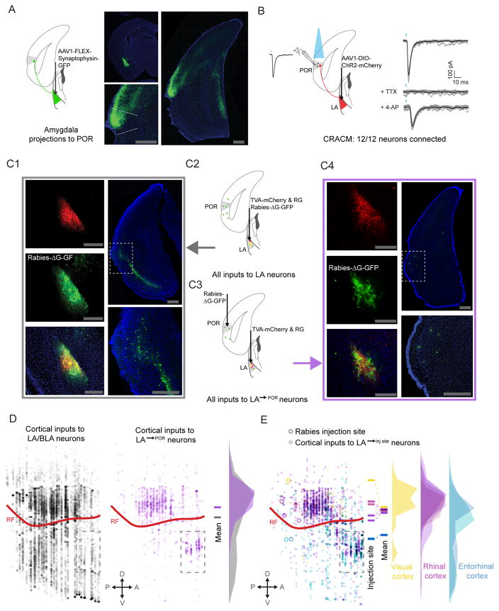
Figure 3. Reciprocal excitatory connectivity between POR and LA.
A. Anterograde viral tracing, using cre-dependent AAV-synaptophysin-GFP, demonstrated dense input from LA to POR.
B. In vitro ChR2-assisted circuit mapping (CRACM) demonstrated a strong, functional excitatory connection from LA to L2/3 pyramidal neurons in POR (n=12/12). TTX (1 μM) and 4-AP (100 μM) were added to the bath solution in order to confirm monosynaptic connectivity.
C. Rabies-based retrograde tracing was used to characterize inputs to glutamatergic LA neurons, and specifically to LA→POR neurons. TVA and rabies glycoprotein were selectively expressed in glutamatergic cells in LA (C1, top left) and G-deleted rabies virus was then injected into either LA (C1, middle and bottom left) or POR, causing LA→POR neurons to selectively be infected (C3 and C4, middle left and bottom left). Rabies-tracing (C2) of inputs to all glutamatergic LA neurons showed strong input from many areas in lateral cortex (C1, right). Projection-specific rabies tracing (C3–C4) of inputs specific to LA→POR neurons also revealed inputs from lateral cortex neurons. Many of these inputs appeared to be from rhinal cortices (C4, right), suggesting a disynaptic, reciprocal excitatory loop from POR to LA and back to POR.
D. Using multi-synapse rabies tracing and whole-brain reconstruction and alignment methods, we confirmed that LA neurons that project to POR received input from a narrower band of neurons in cortex (purple discs), with the greatest density just above the rhinal fissure, in rhinal cortex. Rabies tracing of inputs to all glutamatergic LA neurons (C1–2) demonstrated a larger number and broader distribution of cortical input neurons (gray discs), although the greatest density was still in rhinal cortex.
E. LA projections to different targets in lateral cortex received greater input from cortical regions near the target, suggesting the presence of local, disynaptic reciprocal loops in cortex. Neurons in the immediate vicinity of the LA (dashed rectangle below rhinal fissure) were excluded from analyses in D and E. POR: postrhinal cortex; LA: lateral amygdala; BLA: basolateral amygdala; V1: primary visual cortex; LEnt: lateral entorhinal cortex; rf: rhinal fissure; scale bar: 500 μm. See also Figure S5.

