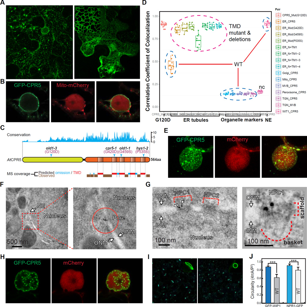
Figure 1. CPR5 Is a Transmembrane Protein Enriched in the Nuclear Pore.
(A and B) Subcellular localization of GFP-tagged CPR5 when transiently expressed in N. benthamiana. Wild-type (WT) CPR5 (A, left), the G420D mutant (A, right), and WT CPR5 co-expressed with a mCherry-tagged marker labeling mitochondria and nucleoplasm (B) were shown. Images were obtained 24 hrs post Agrobacterium infiltration.
(C) CPR5 contains an evolutionarily conserved transmembrane (TM) region at the carboxyl terminus. Top, amino acid conservation map derived from multiple sequence alignments of CPR5 proteins from Micromonas pusilla, Chlorella variabilis, Physcomitrella patens, Sorghum bicolor, Selaginella moellendorffii, Vitis vinifera, Populus tricocarpa, Ricinus communis, Oryza sativa, Zea mays and Arabidopsis thaliana. Middle, schematic of AtCPR5 domain structure with transmembrane TM domains (TMDs) predicted by TMpred. Arrowheads indicate sites of loss-of-function missense mutations. Bottom, LC-MS/MS peptide coverage in the C-terminal half of AtCPR5 purified from transgenic Arabidopsis. Predicted omissions were calculated by PeptideMass for trypsin digestion (see Figure S1C).
(D) Pearson’s correlation coefficients of co-localization between CPR5 and endomembrane organelle markers. WT CPR5 (blue dashed circles) co-localized with both the nuclear envelope (NE) marker (WIT1) and ER-associated granules (see Figure S1B). CPR5 with missense mutations in the TMDs (Mut) and sequential truncations of individual TMDs (N+TM) all exclusively localized in tubular ER structures (magenta dashed circle). TGN (early endosome) and MVB (late endosome) markers were used as a negative control (nc).
(E) Three-dimensional image reconstruction of the nuclear surface in a GFP-CPR5 expressing cell. The nucleoplasm is labeled by free mCherry. Arrowheads indicate large ER-associated granules close to the nuclear surface.
(F and G) Immunoelectron microscopy and tomography analyses of GFP-CPR5 in root cells of transgenic Arabidopsis. Immunogold particles (arrowheads) labeled NPC1 but not NPC2 as antibodies detect only surface-exposed epitopes (G, left). The scaffold and nuclear basket of NPC1 were recognized together with two GFP-CPR5 specific immunogold particles in a projection of the tomographic volume (G, right). ONM/INM, outer/inner nuclear membrane.
(H) Hypolobulated NE and inner nuclear speckles resulted from prolonged overexpression of GFP-CPR5 (40 hrs after Agrobacterium infiltration). (I and J) Nuclear morphology in WT and cpr5 mutant plants. Epidermal cells of 5-day-old seedlings expressing the NE marker GFP-WIP1 were imaged (I). Quantification of the nuclear circularity was performed using GFP-WIP1 and NPR1-GFP as NE and nucleoplasm markers, respectively (J). Data are presented as mean ± SDM (n = 30 cells for each marker and genotype). Asterisks indicate significance (Student’s t-test, ***p-value < 0.001).
See also Figure S1.

