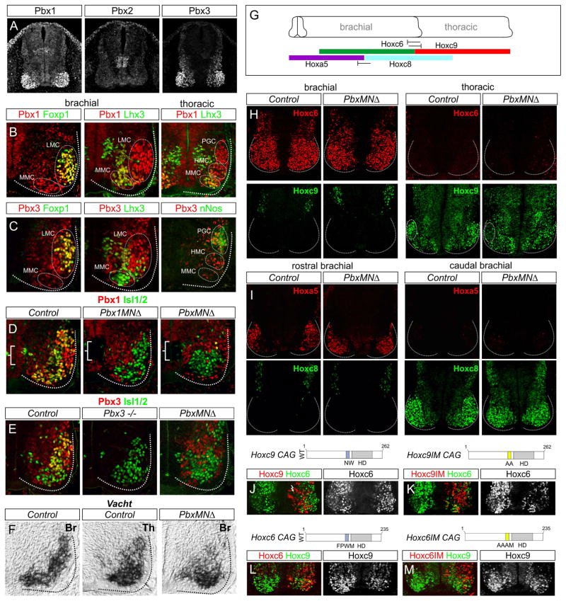
Figure 1. Pbx Genes are Not Required for MN Generation or Establishing Hox Boundaries.
(A) Pbx protein expression in mouse spinal cord at e11.5. Pbx1 is expressed in progenitors and postmitotic MNs. Pbx2 is expressed at low levels in progenitors and postmitotic neurons, and a population of interneuron progenitors. Pbx3 is expressed by postmitotic spinal neurons. (B) Pbx1 colocalizes with Foxp1+ LMC neurons at brachial levels, Lhx3+ MMC neurons, and thoracic HMC and PGC neurons. (C) Pbx3 is restricted to rostral brachial Foxp1+ LMC neurons and excluded from Lhx3+ MMC neurons. At thoracic levels Pbx3 is expressed in HMC and nNos+ PGC neurons. (D) Pbx1 expression is lost in progenitors (brackets) and postmitotic MNs in Pbx1MNΔ and PbxMNΔ mice. (E) Pbx3 expression is lost in Pbx3−/− and PbxMNΔ mice. Pbx3 staining in PbxMNΔ section is from the Pbx3 conditional allele. (F) Vacht mRNA expression at e12.5 in control brachial (Br) and thoracic (Th) MNs and in Br MNs of PbxMNΔ mice. Loss of Br MNs in Pbx mutants does not appear to be due to increased apoptosis (Figure S1H). (G) Summary of Hox expression boundaries in MNs at brachial and thoracic levels. (H) Hoxc6 and Hoxc9 boundaries are maintained in PbxMNΔ mutants, but Hoxc9 levels are reduced in PGC neurons. (I) Hoxa5 and Hoxc8 boundaries are maintained in PbxMNΔ mutants. (J, K) Misexpression of Hoxc9 or a Hoxc9-Pbx interaction mutant (Hoxc9IM) at brachial levels represses Hoxc6. (L, M) Misexpression of Hoxc6 or a Hoxc6IM at thoracic levels represses Hoxc9. See also Figure S1.

