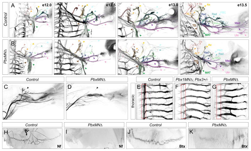
Figure 3. Motor Axon Projections in PbxMNΔ Mice.
(A,B) Dorsal view of forelimb innervation in control and PbxMNΔ mice between e12.0 and e13.5. PbxMNΔ mice have thinner axons and display defects in axonal branching and nerve trajectories. Color coding: Suprascapular Nerve (N.) (Ss, red), Anterior Lateral Thoracic N. (ALT, orange), Axillary N. (Ax, Yellow), Musculocutaneous N. (Mus, neon green), Radial N. (Rad, blue), Posterior Brachial Cutaneous N. (PBC, aqua), Radial/Musculospiral N. (Rad, dark blue), Median N. (Med, dark purple) Ulnar N. (Uln, light purple), Thoracodorsal nerve to Lattismus Dorsi (LD) (TD, brown) and Medial Anterior Thoracic N. to the Cutaneous Maximus (CM) (MAT, dark green). (C,D) In PbxMNΔ; Hb9::GFP mice the dorsal tibialis anterior nerve fails to form in the hindlimb. (E–G) Motor axon projections at thoracic levels showing loss of sympathetic chain ganglia innervation (outlined in red). Mice retaining one allele of Pbx3 also display projection defects. Projections along the intercostal nerves are retained in Pbx mutants, although some aberrant branching is observed. (H,I) Diaphragm innervation defects in PbxMNΔ mutants at e16.5. Motor axons are labeled using Neurofilament (Nf) staining. In PbxMNΔ mice phrenic axons fail to innervate the diaphragm. (J,K) Staining with α-bungarotoxin (Bgt) showing acetylcholine receptor (AChR) clustering in control animals and absence of concentrated clusters in PbxMNΔ mice. See also Figure S3.

