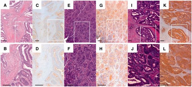
Figure 1.
Aleuria aurantia lectin (AAL) staining in colorectal cancer tissue. Representative microphotographs of weak (A–D), positive (E–H), and high expression (I–L) of AAL. Immunohistochemical staining for AAL was performed using a specific biotinylated AAL. A, B, E, F, I, J) Hematoxylin and eosin staining. C, D, G, H, K, L) Immunostaining for AAL. B, D, F, H, J, L) Representative areas from (A, C, E, G, I, K, and H), respectively. Scale bar = 200 μm.

