Fig. 2.

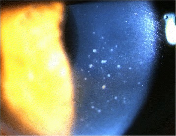
Case 2: slit lamp photograph showing diffuse mutton-fat keratic precipitates within the central area of the cornea. There was mild inflammation in the anterior chamber

Case 2: slit lamp photograph showing diffuse mutton-fat keratic precipitates within the central area of the cornea. There was mild inflammation in the anterior chamber