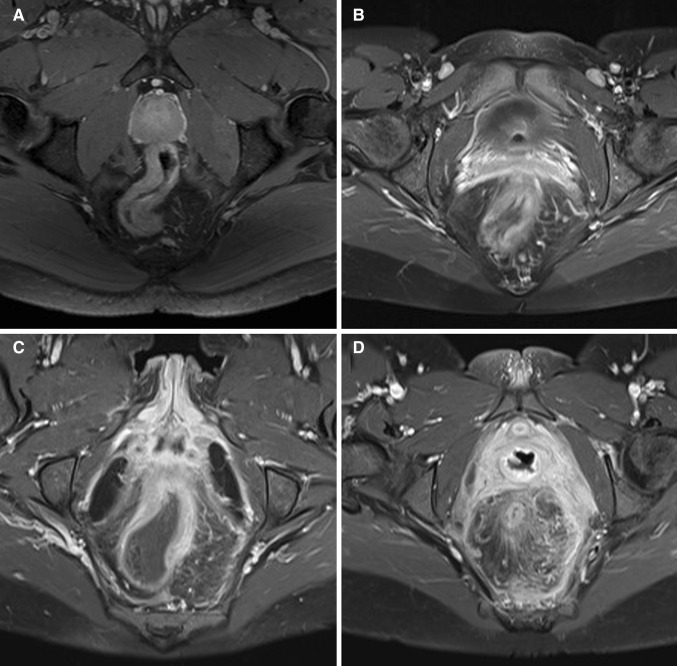
Fig. 1.
Axial oblique fat-saturated post-contrast T1-weighted images of four different patients with Crohn’s disease with different degrees of perimural enhancement. A Equivalent to normal fat tissue. B Minor enhancement. There is blurred demarcation of the bowel wall with or without mild increase of perimural fat tissue signal. C Moderate enhancement. Increase of perimural fat tissue signal but less than nearby vascular structures. D Marked enhancement. Perimural fat tissue signal approaches that of nearby vascular structures. Mesorectal fascia enhancement can be noted

