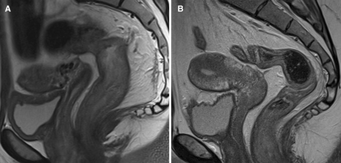
Fig. 3.
Sagittal T2-weighted image of two different patients with Crohn’s disease. A A 25-year-old female with ulcerative proctitis at endoscopy. The image shows increased amount of mesorectal fat tissue (creeping fat) and a subtle increase of perimural vascularity (‘comb sign’) in addition to rectal wall thickening. B A 24-year-old female with no signs of proctitis at endoscopy. There is no increased amount of mesorectal fat tissue and the rectum shows no abnormal MRI features

