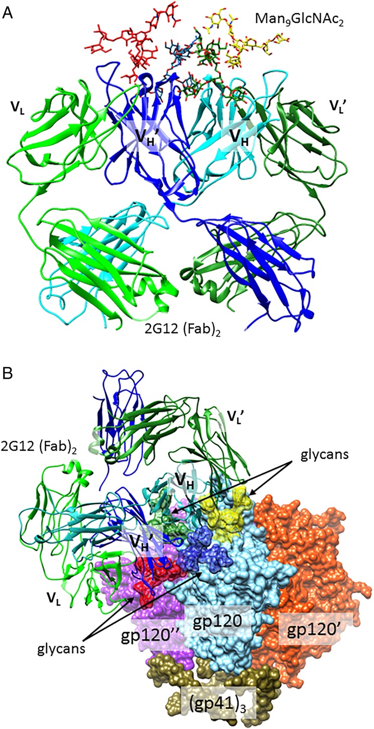
Fig. 2.
2G12 contacts to gp120 glycans. (A) The co-crystal structure of 2G12 Fab dimer in complex with four Man9GlcNAc2 glycans (Calarese et al. 2003; PDB 1OP5). (B) A model of the 2G12 Fab dimer interaction with Man9GlcNAc2 glycans on the surface of trimeric HIV envelope. Glycans at N295, 339, 392 and 332 are depicted in red, blue, yellow and forest green, respectively. All other glycans are deleted for clarity. Although one 2G12 Fab dimer binds to each of the three gp120 protomers, only one 2G12 is shown, for clarity. This model was constructed by aligning the 2G12 (PDB 1OP5) and Env (PDB 4NCO) structures as in Murin et al. (2014). This figure is available in black and white in print and in color at Glycobiology online.

