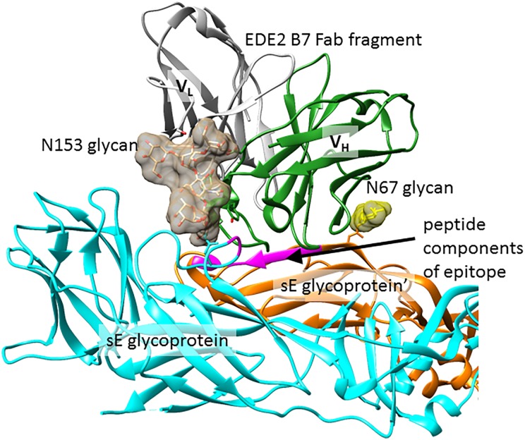
Fig. 5.
Dengue glycoprotein recognition by bnAb EDE2 B7. A crystal structure of dimeric sE glycoprotein in complex with a Fab fragment of bnAb EDE2 B7 (PDB ID: 4UT6). N153 and N67 glycans are shown as charcoal and yellow surfaces, respectively, and peptide components of the epitope are colored purple in both sE glycoprotein chains. The other half of this complex (not shown) is related by C2 symmetry and contains a second Fab fragment making identical contacts to the glycoprotein. This figure is available in black and white in print and in color at Glycobiology online.

