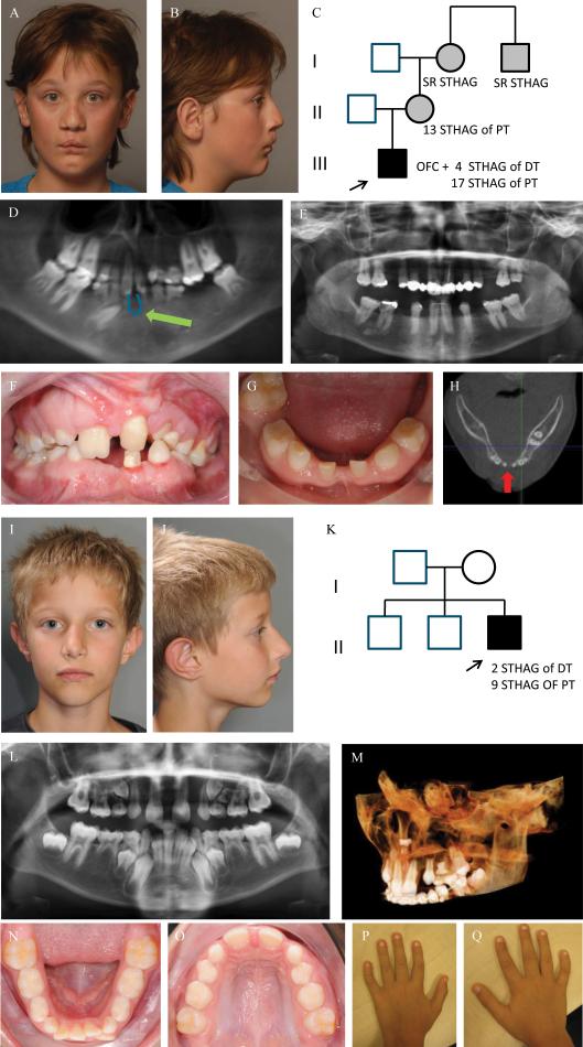
Figure 1. Clinical photographs, orthopantomogram (OPT), image from Cone Beam Computer Tomogram (CBCT) and pedigree of index patient 1 (A-H) and of index patient 2 (I-Q).
Frontal and lateral facial photographs of index patient 1 at 12 yrs of age (A-B), showing a repaired bilateral cleft lip with a left-sided cleft alveolus and a complete cleft of the anterior and posterior palate. He has a wide nasal base, full nasal tip, wide nasal bridge (H) and has a dip in the chin (A, B). His mother, maternal grandmother and her brother also have TA, but no other orofacial abnormalities (C). The patient presents with severe TA or oligodontia: he had agenesis of 4 deciduous teeth (#52, #62, #72 and #82) and misses 18 teeth in the permanent dentition, 17 due to tooth agenesis (#15, #14, #13, #12, #22, #23, #24, #25, #35, #34, #33, #32, #31, #41, #42, #44, #45) (excluding third molars) and 1 (#36) due to extraction. (D). He has a small median mandibular cleft, which can be seen on the orthopantomogram (OPT) at the green arrow (D), the intraoral photographs (F,G) and on the horizontal tomographic view of a CBCT (H). The OPT of the boy's mother shows severe TA, as she misses 13 permanent teeth.
Frontal and lateral photographs of the index patient 2 at 9 yrs of age (I-J) showing mild facial dysmorphic features including a narrow nasal ridge, posteriorly rotated ears with a thin helix, small earlobes, and a long superior crus antihelix. He has unaffected parents and two unaffected brothers (K). The OPT shows tooth agenesis (TA) of two deciduous teeth (#52, #62) and of nine permanent teeth (#17, #15, #14, #12, #22, #25, #27, #35, #45) (L). There is an ectopic tooth germ in the upper right molar area (#17 or #18) and a horizontally impacted premolar germ (#24) in the left upper quadrant (L-M). The occlusal photograph of the mandibular dental arch in the mixed dentition shows malposition of tooth #32 (N). The shape of the palatal cusps of teeth #16 and #26 are abnormal, making them resemble a second molar on the occlusal photograph of the maxillary dental arch (O). He has clinodactyly of the 5th fingers (P-Q).
Abbreviations used: STHAG, selective tooth agenesis; DT, deciduous teeth; PT, permanent teeth

