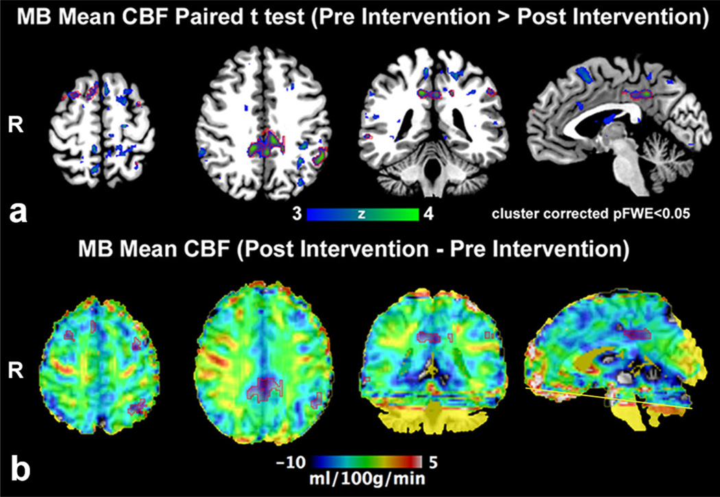
Fig. 3.
(a) Paired analysis (Pre intervention > Post Intervention) of absolute mean CBF (pCASL fMRI) demonstrates larger clusters prior to methylene blue (MB) administration (n = 13); significant clusters are outlined by red ROI. (b) Post intervention minus Pre intervention group absolute mean CBF average demonstrates that the significant clusters belong to the posterior cingulate/precuneus, left inferior parietal lobule and prefrontal cortex consistent with a visuomotor network where CBF is greater prior to MB intervention. Mild CBF increase in the right motor cortex did not achieve significance. Artifact limits evaluation adjacent to the inferior imaging plane (yellow line)

