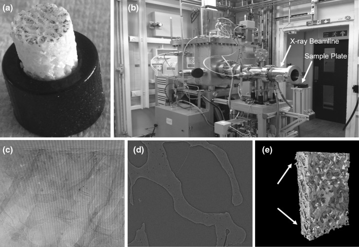
Fig. 1.
Synchrotron micro-CT scan of a trabecular core. a trabecular core mounted into a 3D-printed holder. b X-ray source. c Raw tomography projections. d Reconstructed CT-slice with 1.3 μm/voxels. e 3D model of entire bone core with artifactual drilling damaged on the edge (white arrow) and a synchrotron-scanned region shown in yellow which is far away from the drilling zone

