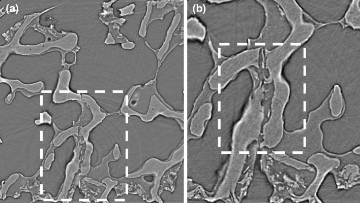
Fig. 6.
Two different spatial resolution scans from I12 beamline at Diamond Light Source, a module 3 scan (3.2 μm/voxel) can only show a general view of the trabecular structural at the same position with precracked three-point bending experiment, b module 4 scan (1.28 μm/voxel) clearly shows a precracked on the bone sample, while dashed sector in (a) can be dearly visualized in (b). However, module 4 takes 60 min per stack, and it is not recommended here as the sample will experience long-time exposure at each loading stage, in which the radiation damage will change the chemical component of the bone and affect the mechanical properties of the experiment

