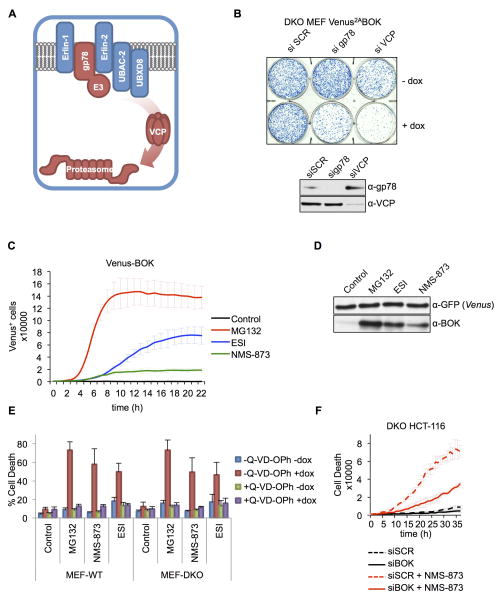
Figure 4. ERAD disruption triggers BOK-induced apoptosis.
A. Schematic representation of ERAD components regulating BOK degradation. B. Clonogenic survival of bax−/−bak−/− MEFs expressing Venus2ABOK transfected with scrambled (SCR), gp78-, or VCP-targeting siRNA and treated with dox for 24 h. C. IncuCyte quantification of Venus-BOK in bax−/−bak−/− MEFs treated with dox and MG132 (10 μM), ESI (1μM), and NMS-873 (10μM) in the presence of 40 μMQ-VD-OPh. D. Immunoblot of Venus and BOK. bax−/−bak−/− MEFs expressing Venus2ABOK were treated as in (C). E. Flow cytometry quantification of Annexin V–stained WT and bax−/−bak−/− MEFs expressing Venus2ABOK and treated with dox and MG132 (10 μM), ESI (1 μM) or NMS-873 (10 μM) for 8 h (mean ± SD of 3 independent experiments). F. IncuCyte quantification of SYTOX Green–stained bax−/−bak−/− HCT116 cells treated with scrambled (SCR) or BOK-targeting siRNA and 10 μM NMS-873. IncuCyte data represented as mean of triplicate samples ± SD and representative of 3 independent experiments (see also Figure S4).

