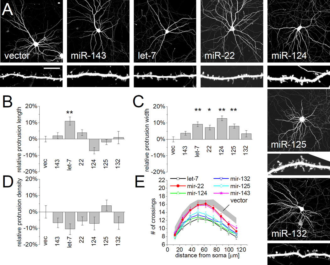
Figure 4. Sponging of FMRP-associated miRNAs differentially affects dendritic spine morphology.
(A) Hippocampal neurons were cotransfected (DIV14+3) with EGFP and sponges for specific miRNAs (see Supplemental Figure S1B, C) to sequester endogenous miRNA. Scale bars represent 10 µm (upper and lower panels). The length (B), width (C) and density (D) of dendritic protrusions was manually quantified. Data was normalized to neurons expressing empty vector (vec). n = 23 to 50 cells each group. Statistical analysis by one-way ANOVA with Dunnett’s post test: * p < 0.05, ** p < 0.01. (E) Sholl analysis of sponge-transfected neurons, measuring the number of dendrites crossing concentric circles at the indicated distance around the cell body. Gray corridor represents neurons transfected with empty vector (mean +/− SEM). n = 40 to 71 neurons each group. Sponge transfected cells are significantly different from control neurons (Two-way ANOVA, p<0.05) for: miR-124 and let-7 (from 37.5 µm radius), miR-125 and miR-132 (from 63.5 µm), miR-22 and miR-143 (from 75 µm).

