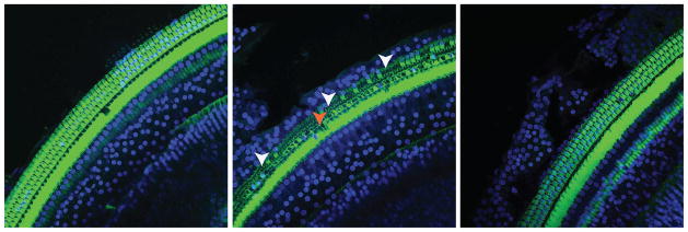
Figure 4.
shows the organ of Corti of chronically implanted rats harvested at Day 28. Stereocilia bundles of HCs were stained with phalloidin-FITC (green) and the nuclei with DAPI (blue). A) shows the organ of Corti of a hypothermia-treated cochlea undergoing EIT. B) shows an EIT normothermic cochlea that had significant outer HC loss (OHC) near the electrode insertion site indicated by the arrows. C) shows a control, non-operated contralateral cochlea.

