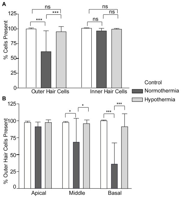
Figure 5.
A) shows the hair cell count comparison between outer and inner normothermia treated, hypothermia treated and control cochleae harvested 28 days post implantation. There was no significant inner hair cell loss, but we observed a pronounced outer hair cell loss in normothermic cochleae. B) Quantification of the total number of outer hair cells (OHCs) present at the end of chronic experiment in basal, middle and apical turn regions of the cochleae in control, normothermic and hypothermia-treated implanted cochleae. Note the significant loss of OHCs in the middle (*P≤0.05) and in the base (***P≤0.005) of the normothermic cochlea. Error bars are S.D.

