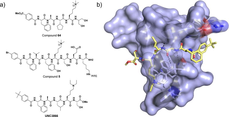
Figure 1. Peptidomimetic chromodomain ligands.

a) Chemical structures of peptidic chromodomain inhibitors. b) Co-crystal structure of chemical probe UNC3866 bound to CBX7 (pdb 5EPJ). The protein surface is shown in light blue and UNC3866 in yellow. Hydrogen bonds between the protein and UNC3866 are shown by a black dotted line.
