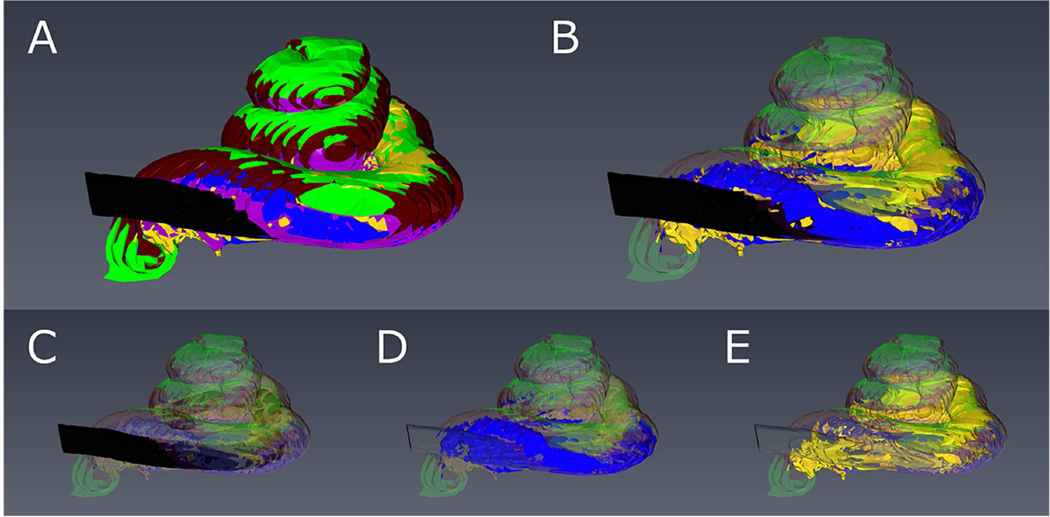
Figure 2.
3-D reconstruction of the implanted right cochlea of Case 17 showing scala tympani (purple), scala media/vestibuli (green), spiral ligament (brown), electrode (black), new bone (blue), and new fibrous tissue (yellow).
A: All elements are shaded. B: Electrode, new bone and fibrous tissue are shaded, and the others are transparent. C: Only the electrode is shaded. D: Only new bone is shaded. E: Only new fibrous tissue is shaded.

