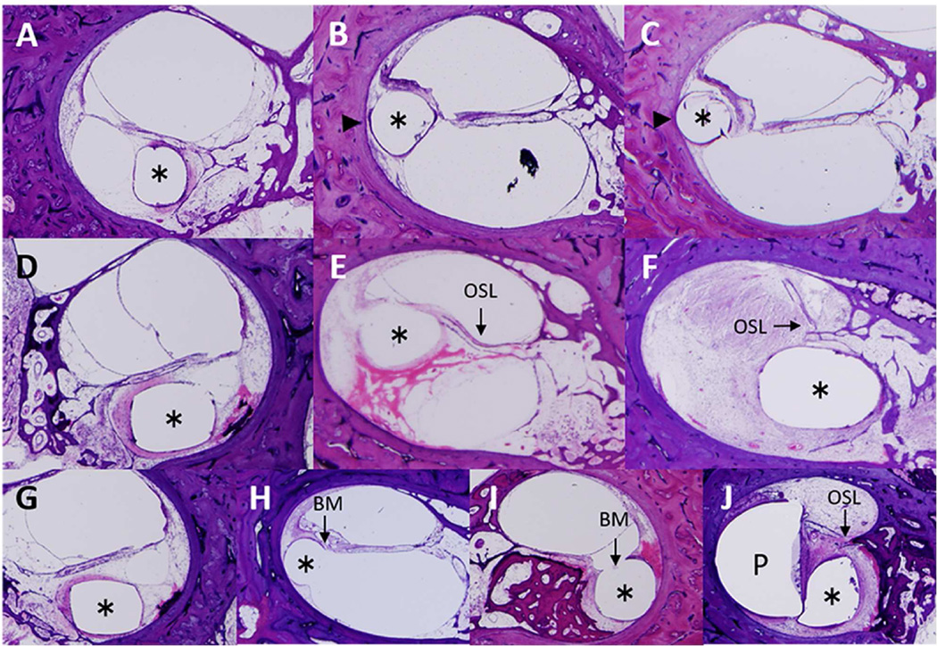
Figure 3.
Quantification and representative images of damage to the lateral cochlear wall, osseous spiral lamina and basilar membrane caused by the electrode. Asterisk indicates the track of the electrode in all ten images. LCW, lateral cochlear wall; OSL, osseous spiral lamina; BM, basilar membrane.
Lateral cochlear wall A: Damage score 0: No dissection into the spiral ligament as seen in Case 9.
B: Damage score 1: Dissection into the spiral ligament (arrowhead) as seen in Case 14.
C: Damage score 2: Dissection into the spiral ligament and contact with the bony lateral cochlear wall (arrowhead) as seen in Case 14.
Osseous spiral lamina D: Damage score 0: Neither displacement, fracture nor dislocation of the osseous spiral lamina as seen in Case 17.
E: Damage score 1: Displacement of the osseous spiral lamina (arrow) as seen in Case 7.
F: Damage score 2: Fracture and dislocation of the osseous spiral lamina (arrow) as seen in Case 15.
Basilar membrane G: Damage score 0: No displacement of the basilar membrane as seen in Case 17.
H: Damage score 1: Displacement of the basilar membrane (arrow) as seen in Case 13.
I: Damage score 2: Disruption of the basilar membrane (arrow) as seen in Case 10.
J: Damage score 2: Loss of the basilar membrane (P: positioner) as seen in Case 1.

