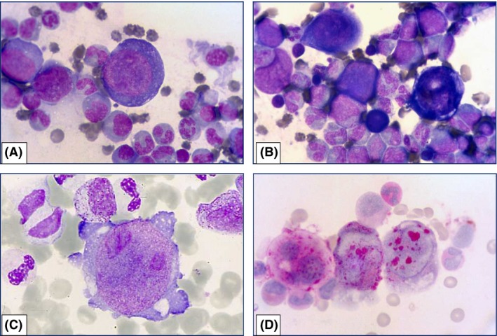
Figure 1.

(A–C) Bone marrow smear showing normal granulopoiesis, lack of maturing erythroblasts, giant proerythroblasts with hyperbasophilic cytoplasm (MGG, 1A,B, x1000), cytoplasmic vacuoles and blebs, and very large nuclear inclusions (MGG, 1C, x1600). (D) Two erythroblasts (right) showing strong PAS positivity with coarse blocks of glycogen; this positivity pattern is different from that observed in the megakaryocyte (left) (PAS reaction, x1000).
