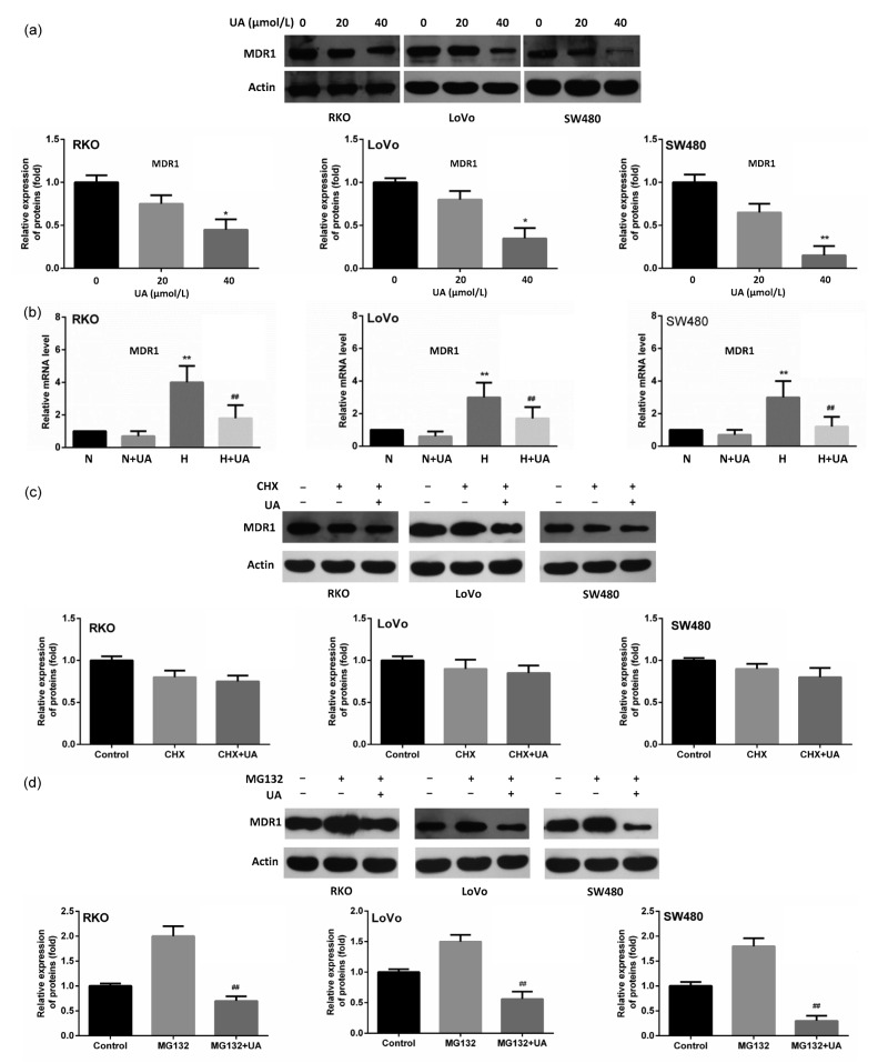
Fig. 3.
Inhibitive effect of UA on MDR1 expression under hypoxia
(a) Cells were cultured under hypoxia and were treated with UA (0, 20 and 40 μmol/L, respectively) for 24 h. MDR1 expression was detected using immunobloting. * P<0.05, ** P<0.01, compared to 0 μmol/L UA group. (b) Cells were treated with 20 μmol/L UA under normoxic and hypoxic conditions and MDR1 mRNA was examined by quantitative RT-PCR. ** P<0.01, compared to normoxia; ## P<0.01, compared to hypoxia. (c) Cells were treated with 50 μg/ml of cycloheximide (CHX) for 24 h, followed with subsequent treatment of UA (20 μmol/L) for another 24 h. MDR1 abundance was examined in all three cell lines using Western blot. (d) Cells were treated with 100 μmol/L of MG132 for 24 h, followed with subsequent treatment of UA (20 μmol/L) for another 24 h. MDR1 expression was detected using Western blot. Data are expressed as mean±SD of triplicate experiments. ## P<0.01, compared to MG132 group

