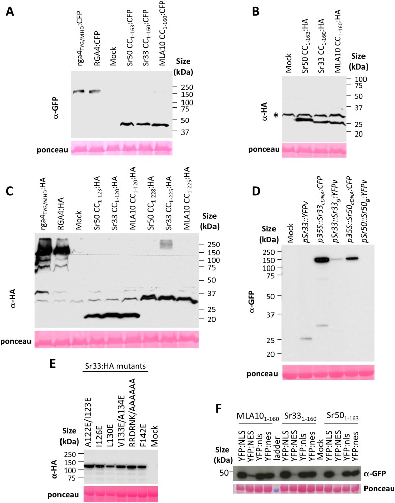
Fig. S3.
(A–F) Immunoblotting showing expression of HA-, CFP-, or YFP-fused proteins. The indicated proteins were extracted from transiently transformed N. benthamiana leaves 24 h after infiltration and were analyzed by immunoblotting with anti-GFP or anti-HA antibodies. Ponceau staining of the large RuBisCO subunit was used to verify equal protein loading. An asterisk marks unspecific background.

