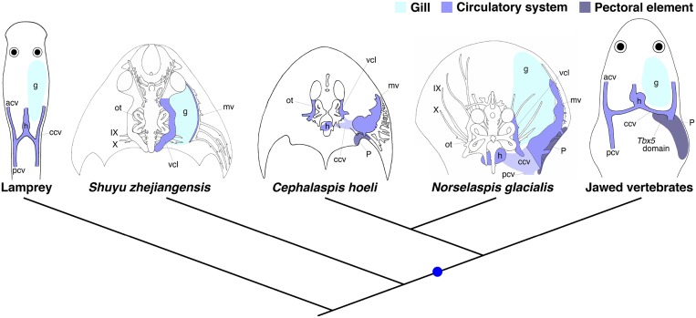
Fig. S10.
Vertebrate phylogeny and evolution of pectoral elements. Head and anterior trunk regions, together with internal anatomy, are schematized in lamprey, galeaspids (Shuyu zhejiangensis), osteostracans (Cephalaspis hoeli and Norselaspis glacialis), and jawed vertebrates. The heart, lateral head vein, and marginal vein in fossil species are reconstructed based on the ossified capsule and canals. The common cardinal vein and posterior cardinal vein of osteostracans are inferred based on the morphology of the pericardial capsule, lateral head and marginal vein canals, and larval lamprey anatomy (41, 52, 53). Osteostracans possess the pectoral elements that are morphologically comparable to that of placoderms, a paraphyletic assemblage of stem-jawed vertebrates (7–12, 40, 42–45). Those components, the pericardial capsule and pectoral elements, are missing in galeaspids, a sister group of osteostracans and jawed vertebrates (7, 10, 12, 40). Note that the topographical relationship of the pectoral appendage in osteostracans relative to the heart, gills, and ccv is comparable to that of jawed vertebrates. Galeaspids was redrawn from ref. 54, and osteostracans was redrawn from refs. 8, 10, 12, and 41. Blue spot indicates gain of pectoral elements, and hypothesized gain of Tbx5 expression posterior to the heart and CNS12. acv, anterior cardinal vein; ccv, common cardinal vein; g, gills; h, heart; IX, glossopharyngeal nerve (ninth cranial nerve); mv, marginal vein; ot, otic components; p, pectoral element; pcv, posterior cardinal vein; vcl, vena capitis lateralis (lateral head vein); X, vagus nerve (10th cranial nerve).

