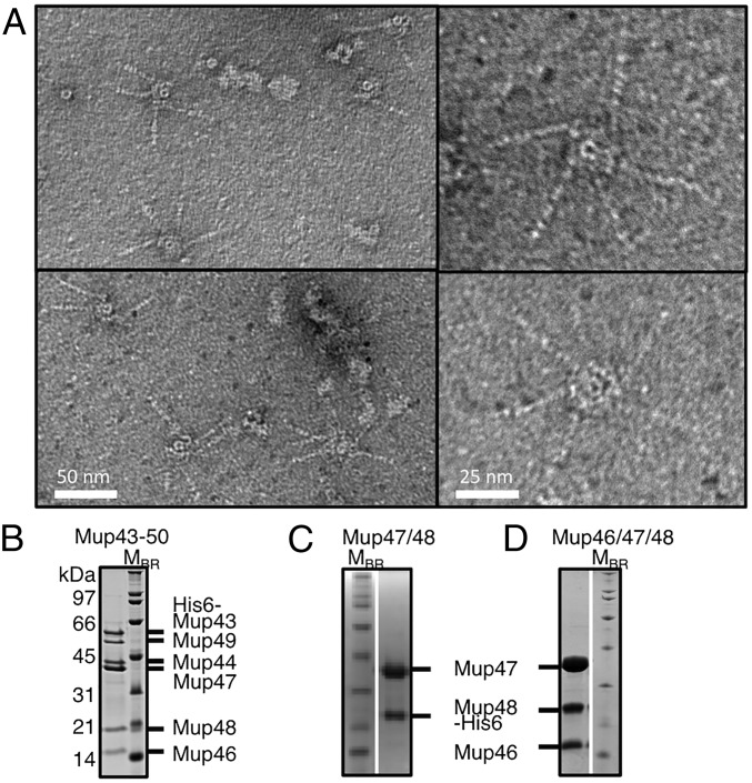
Fig. 2.
Purification of baseplates with attached fibers and assembly intermediates. (A) Representative electron micrographs of purified Mup43–50 complexes, which include tail fibers. (B) The peak fraction of the HM mass complex of Mup43–50 isolated by SEC was analyzed by SDS/PAGE. The Mup43–50 complex was purified using Ni-affinity chromatography by using an N-terminally His6-tagged Mup43 then fractionated by SEC (Fig. S3G). SDS/PAGE gels are shown of the Mup47/Mup48His6 complex (C) and the Mup46/Mup47/Mup48His6 complex (D), after Ni-affinity purification and SEC fractionation (see Fig. S5 for extended SEC data).

