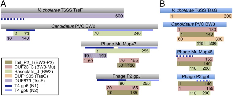
Fig. 5.
Similarities between phage, T6SS, and PVC baseplate wedge proteins. HHpred-detected similarities between BW2 (A) and BW3 (B) proteins and protein families from the Pfam database are shown. Colored rectangles represent regions of similarity with the Pfam family HMMs indicated in the legend. Numbers in the rectangles indicate regions of the given HMM that matched. Hatched rectangles indicate matches with probabilities between 60% and 90%. Nonhatched rectangles represent matches with greater than 90% probability. Blue lines indicate matches to the structure of phage T4 gp6 (PDB ID code 5HX2), with dark blue indicating matches to residues 20–150 and light blue indicating matches to residues 300–450. The proteins shown are examples of TssF (YP_002811746.1), TssG (YP_002811747.1), BW2 (YP_003168848.1), and BW3 (YP_003168847.1) of a PVC, and of E. coli phages Mu and P2 (Datasets S2 and S5).

