Fig. S5.
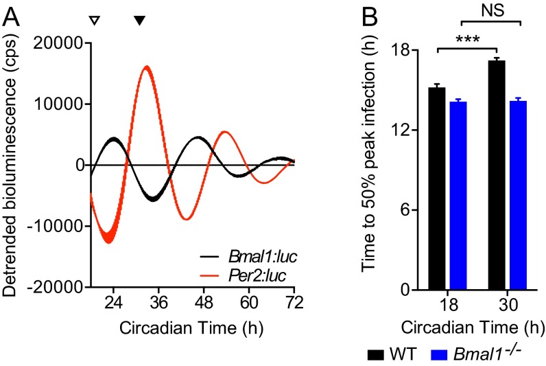
Circadian time effect on MuHV-4 kinetics in WT but not Bmal1−/− cells. (A) Dexamethasone-synchronized mPeriod2:luciferase (Per2:luc) and Bmal1: luciferase (Bmal1:luc) circadian reporter fibroblasts (mean ± SEM; n = 3). Circadian controls for synchronization protocol used in Figs. 4C and 6 C and D. In Fig. 4C, dexamethasone-synchronized WT and Bmal1−/− primary cells were infected with M3:luc MuHV-4 at either CT18 (open arrowhead) or CT30 (solid arrowhead). (B) Kinetic analysis of experiment described in Fig. 4C. Kinetic analysis was performed as shown in Fig. 3B (R2 regression coefficients: WT CT18 = 0.9782, WT CT30 = 0.9932, Bmal1−/− CT18 = 0.9668, Bmal1−/− CT30 = 0.9768). Time to 50% peak infection is significantly decreased in Bmal1−/− cells compared with WT cells [two-way ANOVA (genotype × circadian time of infection): genotype effect, P < 0.0001]. Time-of-day effect on viral replication is observed in WT cells, but not Bmal1−/− cells [time to 50% peak infection two-way ANOVA (genotype × circadian time of infection): post hoc multiple comparisons: NS = not significant, ***P < 0.001].

