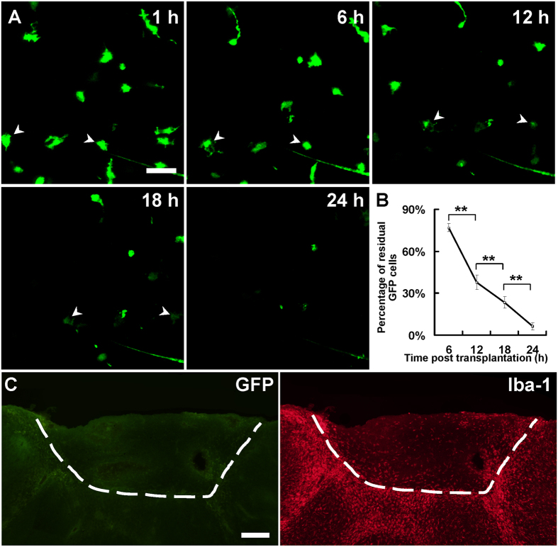
Figure 1. Loss of endogenous microglia in grafted tissue.
(A) In vivo two-photon time-lapse imaging of GFP+ microglia in grafted tissue at 1, 6, 12, 18 and 24 h after transplantation. Most GFP+ cells were lost within 24 h after transplantation. (B) Percentage of residual GFP+ cells in grafted area at 6, 12, 18 and 24 h after transplantation; compared with the number of cells at 1 h. N = 2–3 sections/mouse, 3–6 mice/group, **P < 0.01. (C) An example showing GFP+ endogenous microglia disappeared in grafted tissue at 7 d after transplantation in most cases. Note the host animal was wild-type C57BL/6 mouse and the transplanted tissue was from a CX3CR1GFP/+ mouse. The white dash line shows the boundary between donor and recipient tissue. Scale bar, 25 μm (A); 200 μm (C).

