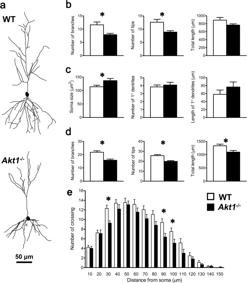
Figure 4. Experiment 4: Examination of the morphological features (mean ± SEM) of pyramidal neurons in the CA1 region of the dorsal hippocampus in female WT mice (n = 13; white bars) and female Akt1−/− mice (n = 11; black bars).
(a) Representative tracings of dorsal hippocampal pyramidal neurons from WT and Akt1−/− mice (scale bar: 50 μm). (b) Apical neuronal properties (left to right: number of branches, number of tips, and total length of the apical tuft (μm)). (c) Basal neuronal properties (left to right: soma size (μm2), number of primary dendrites, and total length of primary dendrites (μm)). (d) Additional basal neuronal properties (left to right: number of branches, number of tips, and total length (μm)). (e) Sholl analysis of basal dendritic complexity within 10-μm concentric spheres around the soma. *p < 0.05.

