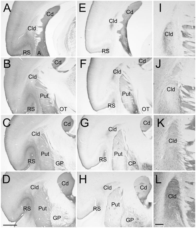
Figure 1. Cytoarchitecture of the tree shrew dorsal claustrum (Cld).

Sections stained with antibodies against calretinin (A-D, and J), substance P (E-H, and I), neuronal nitric oxide synthase (K), and parvalbumin (L) illustrate the location and staining characteristics of the Cld. Sections are all from the same animal. Sections A-D and E-H are arranged from caudal (A, E) to rostral (D, H) at intervals of 600 μm. Sections I-L are adjacent sections through the Cld arranged caudal (I) to rostral (L). A, amygdala, Cd, caudate nucleus, GP, globus pallidus, OT, optic tract, Put, putamen, RS, rhinal sulcus. Scale in D = 1 mm and applies to A-H. Scale in L = 250 μm and applies to I-L.
