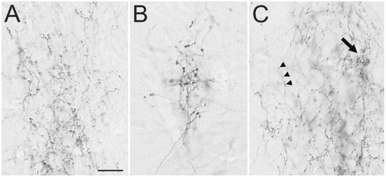
Figure 4. Morphology of V1 projections to the thalamus and claustrum.

Light micrographs illustrate the morphology of axons labeled by an injection of biotinylated dextran amine in V1. A) V1 axons in the dorsal lateral geniculate nucleus form small boutons. B) V1 axons in the ventral pulvinar nucleus form clusters of large boutons. C) V1 axons in the dorsal claustrum form small boutons along thin axons (arrowheads), or dense clusters of slightly larger boutons (arrow). Scale = 20 μm and applies to all panels.
