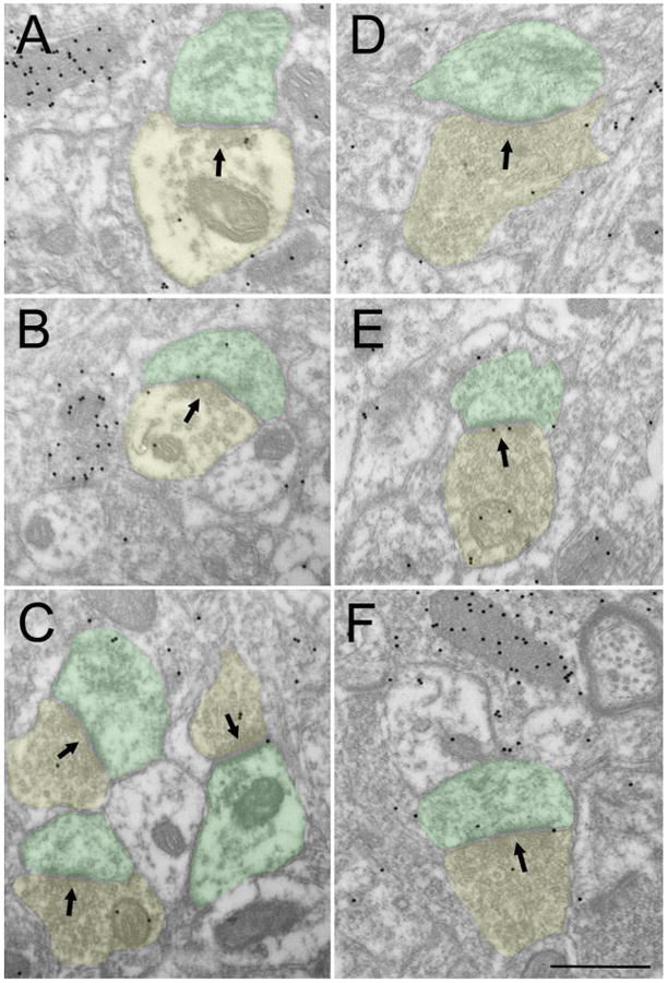
Figure 8. nonGABAergic terminal types in the claustrum.

Electron micrographs illustrate the ultrastructure of nonGABAergic terminals (low density of gold particles, yellow) in the claustrum. NonGABAergic terminals contain sparse (A, B) or dense (C-F) vesicles and primarily contact (black arrows) small nonGABAergic dendrites (green). Scale = 0.5 μm and applies to all panels.
