Figure 6.
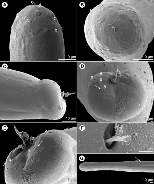
Philometra dispar n. sp. from Carangoides dinema, scanning electron micrographs of male. A, B: Cephalic end, dorsoventral and apical views, respectively. C, D: Caudal end, lateral and apical views, respectively (arrow indicates phasmid). E: Caudal end, subdorsal view (arrows indicate phasmids). F: Deirid. G: Anterior end of body, dorsoventral view (arrow indicates location of deirid). Abbreviations: a, amphid; b, submedian pair of cephalic papillae of external circle; c, submedian cephalic papilla of internal circle; d, lateral cephalic papilla of internal circle; e, caudal papillae in region of cloacal aperture; f, caudal papilla of last postanal pair; g, caudal mound; o, oral aperture.

